
The Rear Organs of the Upper Abdomen Anatomy Model Labelled is a helpful learning tool for students who want to understand what sits behind the front abdominal organs. Many anatomy lessons focus on the stomach, liver, and intestines because they are easy to recognize. But the rear upper abdomen is just as important. It includes organs and structures that support digestion, blood filtration, hormone balance, fluid control, and waste removal.
For students, a labelled anatomy model makes this area easier to study because it turns a complex three-dimensional space into something visual and memorable. Instead of only reading organ names from a textbook, you can see where each organ sits, how it connects to nearby structures, and why its position matters.
Why the Rear Upper Abdomen Matters in Anatomy
The upper abdomen is the area between the chest and the middle part of the belly. When we talk about the rear upper abdomen, we are focusing on organs and structures that sit toward the back of this region, closer to the spine than to the abdominal wall.
This area is not empty space. It contains several important organs, major blood vessels, nerves, ducts, and supporting tissues. Some organs are fully inside the abdominal cavity, while others are partly protected behind the peritoneum, the thin lining that covers many abdominal organs.
A student studying The Rear Organs of the Upper Abdomen Anatomy Model Labelled can better understand how the body is arranged in layers. This is important because anatomy is not just about memorizing names. It is about understanding position, function, and relationships.
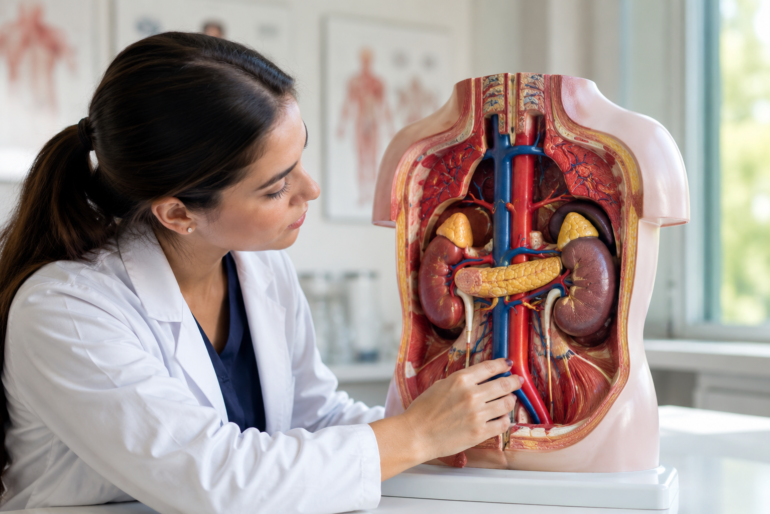
For example, the kidneys are not located in the front of the abdomen. They sit toward the back, on both sides of the spine. The pancreas stretches across the rear upper abdomen, partly behind the stomach. The adrenal glands sit like small caps above the kidneys. These positions affect how doctors examine pain, diagnose disease, and read medical images.
What Does “Rear Organs of the Upper Abdomen” Mean?
The phrase “rear organs of the upper abdomen” usually refers to organs and structures located in the posterior part of the upper abdominal region. In simple words, these are the deeper organs found closer to the back of the body.
In an anatomy model, this region may show:
| Structure | Basic Location | Main Role |
|---|---|---|
| Kidneys | Back of upper abdomen | Filter blood and produce urine |
| Adrenal glands | On top of kidneys | Produce important hormones |
| Pancreas | Behind the stomach | Helps digestion and blood sugar control |
| Spleen | Upper left abdomen, toward the back | Supports immune function and blood filtering |
| Abdominal aorta | Front of spine | Carries oxygen-rich blood downward |
| Inferior vena cava | Right side of spine | Carries blood back to the heart |
| Diaphragm | Above upper abdominal organs | Helps breathing and separates chest from abdomen |
The Rear Organs of the Upper Abdomen Anatomy Model Labelled may also include nearby muscles, nerves, and ducts depending on how detailed the model is.
The Rear Organs of the Upper Abdomen Anatomy Model Labelled: Main Parts Students Should Know
A good labelled anatomy model helps students identify each organ clearly. It may use numbers, color coding, removable pieces, or printed labels to show where each structure belongs.
The Rear Organs of the Upper Abdomen Anatomy Model Labelled usually highlights the organs that are harder to imagine from a flat diagram. Because these organs sit deeper inside the body, students often struggle to picture their exact placement.
Let’s look at the major parts one by one.
Kidneys: The Body’s Natural Filters
The kidneys are two bean-shaped organs located toward the back of the upper abdomen. They sit on either side of the spine, with the right kidney usually a little lower than the left because of the liver’s position.
Their main job is to filter blood, remove waste, balance fluids, and help control blood pressure. They also support red blood cell production through hormones.
In an anatomy model, the kidneys are often shown as reddish-brown organs with a curved outer edge. Some models cut one kidney open to show the renal cortex, renal medulla, renal pelvis, and ureter.
Students should remember these key kidney-related parts:
| Kidney Part | What It Does |
|---|---|
| Renal cortex | Outer filtering area |
| Renal medulla | Inner region with pyramid-shaped structures |
| Renal pelvis | Collects urine before it enters the ureter |
| Ureter | Carries urine from kidney to bladder |
| Renal artery | Brings blood into the kidney |
| Renal vein | Carries filtered blood away |
The National Institute of Diabetes and Digestive and Kidney Diseases describes the kidneys as essential organs for removing waste and extra fluid from the blood. This is why kidney position and function are important in both biology and medical studies.
Adrenal Glands: Small Organs With Big Hormonal Roles
The adrenal glands are small triangular or cap-like glands located on top of each kidney. They may look tiny on a model, but their functions are powerful.
These glands produce hormones such as cortisol, aldosterone, adrenaline, and noradrenaline. These hormones help the body respond to stress, regulate salt and water balance, and support blood pressure control.
In The Rear Organs of the Upper Abdomen Anatomy Model Labelled, the adrenal glands are easy to miss because they are small. Students should look carefully above the kidneys. The right adrenal gland often appears more triangular, while the left one may look more crescent-shaped.
The adrenal glands have two main regions:
| Region | Main Function |
|---|---|
| Adrenal cortex | Produces steroid hormones such as cortisol and aldosterone |
| Adrenal medulla | Produces adrenaline-related hormones |
A useful student tip is to connect the word “adrenal” with “adrenaline.” That makes it easier to remember their role in stress response.
Pancreas: A Deep Organ Behind the Stomach
The pancreas is one of the most important rear upper abdominal organs. It lies behind the stomach and stretches horizontally across the abdomen. It has a head, body, and tail.
The head of the pancreas sits near the duodenum, which is the first part of the small intestine. The tail reaches toward the spleen on the left side.
The pancreas has two major jobs. First, it produces digestive enzymes that help break down food. Second, it produces hormones such as insulin and glucagon, which help regulate blood sugar.
In a labelled anatomy model, the pancreas may look like a long, flat, yellowish organ. It is usually placed behind the stomach, so some models remove the stomach to show it clearly.
The main parts of the pancreas include:
| Pancreas Part | Location or Role |
|---|---|
| Head | Near the duodenum |
| Body | Middle section |
| Tail | Extends toward spleen |
| Pancreatic duct | Carries digestive enzymes |
| Islets of Langerhans | Produce insulin and glucagon |
Students often confuse the pancreas with the spleen because both are in the upper abdomen. The easiest way to remember the difference is this: the pancreas is long and central, while the spleen is more rounded and sits on the upper left side.
Spleen: Blood Filtering and Immune Support
The spleen is located in the upper left part of the abdomen, tucked under the rib cage and near the stomach. It sits more toward the back than many students expect.
The spleen filters blood, removes old red blood cells, stores platelets, and supports the immune system. It helps the body identify and fight certain infections.
In The Rear Organs of the Upper Abdomen Anatomy Model Labelled, the spleen may appear as a dark red or purple oval-shaped organ. It is often placed near the tail of the pancreas.
The spleen is not part of the digestive tract, even though it is near digestive organs. This is a common student mistake. It belongs more closely to the lymphatic and immune systems.
Important points about the spleen:
| Feature | Student Note |
|---|---|
| Location | Upper left abdomen |
| Shape | Oval or curved |
| Nearby organs | Stomach, pancreas, left kidney |
| Main role | Blood filtering and immune defense |
A helpful memory trick is to associate the spleen with “screening” blood. It screens blood for old cells and helps the immune system respond.
Diaphragm: The Roof of the Upper Abdomen
The diaphragm is a dome-shaped muscle that separates the chest cavity from the abdominal cavity. It sits above the upper abdominal organs and plays a major role in breathing.
When you inhale, the diaphragm contracts and moves downward. This creates space for the lungs to expand. When you exhale, it relaxes and moves upward.
In an anatomy model, the diaphragm may appear as a curved muscular sheet above the liver, stomach, spleen, and kidneys. It is especially important when studying the rear upper abdomen because several organs sit directly below it.
The kidneys, adrenal glands, liver, spleen, and stomach all relate to the diaphragm in some way. This explains why pain from upper abdominal organs can sometimes feel like chest, back, or shoulder discomfort.
Abdominal Aorta: The Main Blood Supply Route
The abdominal aorta is one of the most important blood vessels shown in a rear upper abdomen model. It runs down in front of the spine and carries oxygen-rich blood from the heart to the abdomen, pelvis, and legs.
This vessel gives off branches that supply the stomach, liver, spleen, kidneys, intestines, and other structures. In a model, it is usually shown as a large red vessel.
Students should pay close attention to the renal arteries, which branch from the abdominal aorta and go to the kidneys. These arteries are important because the kidneys need a strong blood supply to filter blood effectively.
Key branches students may see include:
| Branch | Supplies |
|---|---|
| Celiac trunk | Stomach, liver, spleen, pancreas |
| Superior mesenteric artery | Small intestine and part of large intestine |
| Renal arteries | Kidneys |
| Gonadal arteries | Reproductive organs |
| Inferior mesenteric artery | Lower large intestine |
Even if a basic student model does not show every branch, recognizing the abdominal aorta helps students understand how blood reaches the rear upper abdominal organs.
Inferior Vena Cava: The Return Path to the Heart
The inferior vena cava, often called the IVC, is a large vein that carries oxygen-poor blood from the lower body back to the heart. It runs along the right side of the spine and close to the abdominal aorta.
In The Rear Organs of the Upper Abdomen Anatomy Model Labelled, the inferior vena cava is usually shown as a large blue vessel. It receives blood from the kidneys through the renal veins.
Students sometimes confuse the abdominal aorta and inferior vena cava because they sit close together. The easiest difference is color and function. In most models, the aorta is red because it carries oxygen-rich blood. The inferior vena cava is blue because it returns blood to the heart.
| Vessel | Model Color | Main Function |
|---|---|---|
| Abdominal aorta | Red | Carries blood away from heart |
| Inferior vena cava | Blue | Carries blood back to heart |
This simple comparison helps students answer anatomy lab questions quickly.
Retroperitoneal Organs: Why Position Matters
Many rear upper abdominal organs are described as retroperitoneal. This means they are located behind the peritoneum, the lining that covers much of the abdominal cavity.
The kidneys, adrenal glands, most of the pancreas, and major blood vessels are retroperitoneal. This location matters because retroperitoneal organs are deeper and less mobile than organs suspended inside the peritoneal cavity.
A common memory aid for retroperitoneal organs is SAD PUCKER, which includes structures such as the suprarenal glands, aorta and inferior vena cava, duodenum, pancreas, ureters, colon parts, kidneys, esophagus, and rectum.
Students do not need to memorize every advanced detail at once. But they should understand the basic idea: some organs are at the back of the abdomen and sit behind the abdominal lining.
This helps explain why kidney pain is often felt in the back or flank area instead of the front belly.
How a Labelled Anatomy Model Helps Students Learn Faster
An anatomy model gives students something that flat textbook diagrams cannot fully provide: depth. The abdomen is not a simple flat map. Organs overlap, curve, and sit at different levels.
The Rear Organs of the Upper Abdomen Anatomy Model Labelled helps students learn through visual memory. When students can see the kidney behind the stomach area or the pancreas crossing the back of the abdomen, the information becomes easier to retain.
A labelled model also helps with active recall. Instead of reading the label first, students can cover the labels and try to name each structure. This makes study sessions more effective.
Here are practical ways students can use the model:
| Study Method | How It Helps |
|---|---|
| Label covering | Tests memory without hints |
| Side-by-side textbook study | Connects model with diagrams |
| Group questioning | Improves recall through discussion |
| Drawing practice | Builds spatial understanding |
| Function matching | Links organ names with what they do |
For example, a student can point to the pancreas and ask, “What does this organ produce?” Then they can answer, “Digestive enzymes and blood sugar hormones.” This method is better than memorizing isolated facts.
Common Student Mistakes When Studying Rear Upper Abdomen Organs
Students often make predictable mistakes when learning this region. Knowing these mistakes in advance can save time.
One common mistake is thinking the kidneys are located in the lower belly. In reality, they sit higher and toward the back, partly protected by the lower ribs.
Another mistake is assuming the pancreas is in the front of the abdomen because it is often shown clearly in diagrams. In the real body, it lies behind the stomach.
Students also mix up the spleen and pancreas. The spleen is more rounded and located in the upper left abdomen, while the pancreas is longer and stretches across the back of the stomach area.
Some students forget the adrenal glands completely because they are small. But these glands are important, especially in lessons about hormones and stress response.
Common mistakes include:
| Mistake | Better Understanding |
|---|---|
| Kidneys are low in the belly | They sit high and toward the back |
| Pancreas is in the front | It lies behind the stomach |
| Spleen is digestive | It supports blood and immunity |
| Adrenal glands are minor | They control important hormones |
| Aorta and IVC are the same | One carries blood away, the other returns it |
A labelled model helps correct these errors because students can see the positions clearly.
Real Classroom Scenario: Learning With a Labelled Model
Imagine a biology student preparing for a practical exam. The teacher places an abdominal model on the table and asks the student to identify the organ above the kidney.
A student who only memorized textbook notes may hesitate. But a student who has studied The Rear Organs of the Upper Abdomen Anatomy Model Labelled will likely recognize the adrenal gland quickly.
Now imagine the teacher points to the long organ behind the stomach and asks for its function. The student identifies the pancreas and explains that it supports digestion and blood sugar regulation.
This is where models become useful. They prepare students for real identification tasks, not just written definitions.
In medical, nursing, biology, and anatomy classes, students are often tested on both structure and function. A good model supports both.
Best Way to Study the Rear Upper Abdomen for Exams
Studying anatomy works best when students combine visual learning with repetition. Do not simply stare at the model and hope the names stay in your memory.
Start with broad regions. Identify the spine, diaphragm, kidneys, and major vessels first. Then move to smaller structures such as adrenal glands, renal arteries, and pancreatic ducts.
A simple study order could look like this:
- Locate the diaphragm.
- Find the kidneys.
- Identify the adrenal glands above the kidneys.
- Trace the pancreas behind the stomach area.
- Locate the spleen on the upper left side.
- Find the abdominal aorta and inferior vena cava.
- Review blood vessels connected to the kidneys.
- Match each organ with its main function.
This step-by-step method prevents confusion. It also builds a mental map of the area.
For better memory, students can say the organ name, location, and function out loud. For example: “The adrenal glands sit above the kidneys and produce hormones.” Speaking the answer makes recall stronger.
Important Functions of Rear Upper Abdominal Organs
The rear upper abdomen is not just an anatomical location. It is a working center for many body systems.
The kidneys support the urinary system. The adrenal glands support the endocrine system. The pancreas supports both digestion and hormone balance. The spleen supports the immune and blood systems. The diaphragm supports breathing. The abdominal aorta and inferior vena cava support circulation.
This means one region connects several major systems.
| Organ or Structure | Body System | Main Function |
|---|---|---|
| Kidneys | Urinary | Filter blood and make urine |
| Adrenal glands | Endocrine | Produce hormones |
| Pancreas | Digestive and endocrine | Enzymes and blood sugar control |
| Spleen | Lymphatic and immune | Blood filtering and immune support |
| Diaphragm | Respiratory and muscular | Breathing support |
| Abdominal aorta | Circulatory | Blood supply |
| Inferior vena cava | Circulatory | Blood return |
Understanding this table helps students see why anatomy is connected. Organs do not work alone. They support one another.
Why Labels Are Important on Anatomy Models
Labels help students connect visual shape with correct terminology. This is especially useful for beginners who may not yet recognize organs by shape.
However, labels should not become a crutch. The best way to use a labelled anatomy model is to study with labels first, then test yourself without them.
When reviewing The Rear Organs of the Upper Abdomen Anatomy Model Labelled, students should focus on three things:
- Name of the organ.
- Position of the organ.
- Function of the organ.
For example, do not only memorize “kidney.” Learn that the kidneys sit posteriorly on both sides of the spine and filter blood to form urine.
This deeper approach makes the information more useful in exams and real learning.
How This Anatomy Model Supports Medical and Biology Students
Medical students, nursing students, biology learners, and health science students all benefit from anatomy models. A model makes it easier to understand organ relationships before moving into more advanced topics like pathology, imaging, or surgery.
For biology students, the model helps connect body systems. For nursing students, it supports clinical understanding of pain location and organ function. For medical students, it provides a foundation for imaging, diagnosis, and anatomical orientation.
The Rear Organs of the Upper Abdomen Anatomy Model Labelled is especially useful because this part of the body is difficult to understand from surface view alone. You cannot see the kidneys, pancreas, adrenal glands, or major vessels from the outside. A model reveals how these structures sit inside the body.
Simple Tips to Remember Each Rear Upper Abdominal Organ
Students often remember anatomy better when they use simple associations.
For the kidneys, think “filters.”
For the adrenal glands, think “stress hormones.”
For the pancreas, think “digestion and sugar.”
For the spleen, think “blood and immunity.”
For the diaphragm, think “breathing roof.”
For the abdominal aorta, think “main red road.”
For the inferior vena cava, think “main blue return.”
These memory cues are not a replacement for proper study, but they make the first learning stage easier.
You can also create a quick sentence:
“Behind the belly, kidneys filter, adrenals signal, pancreas balances, spleen protects, and vessels carry life.”
It sounds simple, but it gives students a quick mental picture of the rear upper abdomen.
How to Read a Labelled Upper Abdomen Model Correctly
When looking at a labelled anatomy model, always begin with orientation. Ask yourself: Is this a front view, back view, side view, or cutaway view?
A rear organ model may show organs from the front after removing the stomach and intestines, or it may show a posterior view. This can change how structures appear.
Next, identify the midline. The spine, abdominal aorta, and inferior vena cava help you understand left and right placement.
Then look for paired organs. The kidneys and adrenal glands appear on both sides. After that, look for single organs like the pancreas, spleen, and major vessels.
Finally, connect structure with function. That is where real understanding begins.
FAQ About Rear Upper Abdomen Anatomy Models
What organs are usually shown in the rear upper abdomen?
The main organs include the kidneys, adrenal glands, pancreas, spleen, diaphragm, abdominal aorta, and inferior vena cava. Some models may also show the duodenum, ureters, nerves, and muscles.
Why is the pancreas considered a rear upper abdominal organ?
The pancreas lies behind the stomach and stretches across the upper abdomen. Because of its deeper position, it is often studied with posterior or retroperitoneal structures.
Are the kidneys in the upper or lower abdomen?
The kidneys are located in the posterior upper abdomen, on both sides of the spine. They sit higher than many students expect and are partly protected by the lower ribs.
What is the easiest way to study The Rear Organs of the Upper Abdomen Anatomy Model Labelled?
Start with large structures first, such as the kidneys, diaphragm, and major vessels. Then move to smaller parts like adrenal glands, ducts, and blood vessel branches. Test yourself by covering the labels.
Is the spleen part of the digestive system?
No. The spleen sits near digestive organs, but it mainly supports blood filtering and immune function. Its location causes confusion, but its function is not digestive.
Why do students need labelled anatomy models?
Labelled models help students understand organ position, shape, and relationship. They are useful for visual learning, practical exams, and building a clear three-dimensional understanding of the body.
Conclusion
The Rear Organs of the Upper Abdomen Anatomy Model Labelled gives students a clearer way to understand one of the body’s deeper and more complex regions. Instead of memorizing organ names from a flat page, students can see how the kidneys, adrenal glands, pancreas, spleen, diaphragm, abdominal aorta, and inferior vena cava fit together.
This kind of model is especially useful because the rear upper abdomen includes organs from several body systems. It connects digestion, blood filtration, hormone control, breathing, immunity, and circulation in one compact area.
For students, the best approach is simple: learn the location, understand the function, and review the labels until the layout feels natural. When used properly, The Rear Organs of the Upper Abdomen Anatomy Model Labelled becomes more than a classroom object. It becomes a practical study partner that makes anatomy easier, clearer, and more memorable.
Students who want to go deeper can also review basic human anatomy concepts to connect organ structure with body systems and everyday function.