
If you have ever wondered how doctors can “see” what the brain is doing in real time, EEG Tech is a big part of that answer. In hospitals, EEG Tech helps capture the brain’s electrical signals and turn them into information doctors can use to diagnose seizures, evaluate unexplained confusion, guide treatment in the ICU, and support safer clinical decisions. It is not just a test you get once and forget. In many cases, it becomes a continuous window into brain function when symptoms are unclear or when minutes matter.
This article breaks down what EEG Tech does inside hospitals, how the process works from start to finish, what conditions it helps diagnose, and why EEG monitoring is often the difference between guessing and knowing.
What EEG Tech Means in a Hospital Setting
EEG stands for electroencephalography, but in day to day hospital life, people often say “EEG” to mean both the test and the whole workflow behind it. EEG Tech refers to the technology, the methods, and the trained professionals (EEG technologists) who record and support EEG testing.
In a hospital, EEG Tech typically includes:
- EEG machines and amplifiers that capture tiny electrical signals from the scalp
- Electrodes and conductive materials applied to specific scalp locations
- Recording software that displays brainwave patterns and flags events
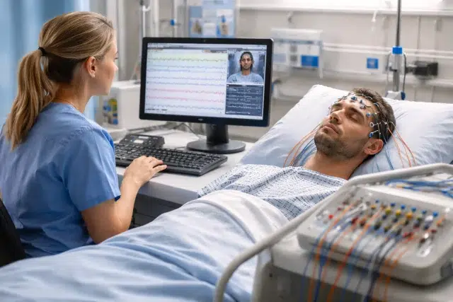
- Video EEG systems that sync brain activity with patient behavior
- Continuous EEG (cEEG) setups used in ICUs for hours or days
- Protocols and standards that help ensure the recording is accurate and clinically useful
Professional guidance and standards for EEG practice and recordings are maintained by organizations such as the American Clinical Neurophysiology Society (ACNS) and the International League Against Epilepsy (ILAE) working with the International Federation of Clinical Neurophysiology (IFCN). These standards help ensure EEG recordings are technically adequate and interpretable in real clinical environments.
Why EEG Tech Matters So Much in Hospitals
Hospitals deal with high stakes situations where symptoms can look similar but mean very different things. A patient might be “out of it,” unresponsive, or having strange movements. Is it a seizure? Medication side effects? Sleep deprivation? Metabolic issues? Stroke? Infection? EEG Tech helps narrow the possibilities by showing whether abnormal electrical activity is happening.
This matters because conditions like epilepsy are common and often underdiagnosed or misinterpreted. The World Health Organization estimates epilepsy affects around 50 million people worldwide, and nearly 80 percent live in low and middle income countries.
In the United States, CDC reporting commonly cites millions living with epilepsy, reinforcing that seizure disorders are not rare and that diagnostic tools like EEG have real public health impact.
How EEG Tech Works: The Process in Plain Language
Even though EEG can look intimidating, the core idea is simple: the brain produces electrical activity, and electrodes placed on the scalp can detect patterns in that activity.
Step 1: Preparing the patient and the scalp
A technologist measures the head and applies electrodes using a standardized placement system. The scalp is prepped to reduce signal noise because EEG signals are extremely small.
Step 2: Recording brain activity
The EEG machine records electrical signals and turns them into waveforms. Depending on the reason for the test, the patient may:
- rest with eyes open and closed
- do deep breathing (hyperventilation)
- see flashing lights (photic stimulation)
- try to fall asleep (for sleep EEG)
- be monitored continuously in the ICU
Step 3: Making sure the recording is high quality
This is where hospital EEG Tech becomes more than “stick on electrodes and press start.” In real life, recordings can be ruined by:
- muscle tension
- movement artifacts
- poor electrode contact
- electrical interference
- sweating, hair products, or patient agitation
A skilled technologist constantly adjusts, troubleshoots, and documents changes so the interpreting physician has clean data.
Step 4: Interpretation and clinical decision making
EEG technologists do not diagnose, but the way they record, annotate events, and maintain quality directly affects diagnostic accuracy. A physician trained in EEG interpretation uses the recording to look for patterns consistent with seizures, epileptiform activity, encephalopathy, sleep disorders, and other brain function changes.
Routine EEG vs Continuous EEG: What Hospitals Use and Why
Not all EEG tests are the same. Hospitals use different types depending on urgency, symptoms, and clinical goals.
Routine EEG (short study)
Often 20 to 30 minutes (sometimes longer), used for:
- suspected seizures
- fainting episodes with unclear cause
- confusion that comes and goes
- baseline brain function after an event
Minimum recording standards and best practices are discussed in international guidance from ILAE and IFCN, supporting consistent, interpretable studies.
Continuous EEG (cEEG)
Hours to days of monitoring, often in ICU, used for:
- unexplained coma or decreased consciousness
- suspected nonconvulsive seizures
- monitoring after brain injury, stroke, or cardiac arrest
- titrating sedatives in status epilepticus treatment
A key reason cEEG is so valuable is that not all seizures look like dramatic shaking. In critical care, nonconvulsive seizures can be missed without EEG monitoring, which is why modern ICU neurology relies heavily on EEG Tech.
The Big Clinical Problem EEG Tech Solves: Nonconvulsive Seizures
One of the most important roles of hospital EEG Tech is detecting seizures that do not have obvious outward signs.
A patient might look:
- sleepy but not waking up
- confused and agitated
- “staring” or not responding
- stable on the outside while the brain is in distress
Research reviews on nonconvulsive status epilepticus (NCSE) report that studies using continuous EEG suggest NCSE can be present in a meaningful range of patients with unexplained coma, with reported ranges in published literature.
That’s why EEG Tech is often treated like a safety net in ICUs. Without it, clinicians can miss ongoing brain seizures that can worsen outcomes if left untreated.
Conditions EEG Tech Helps Diagnose or Evaluate in Hospitals
EEG is best known for seizures, but in hospitals it supports decision making across multiple scenarios.
1) Epilepsy and seizure disorders
EEG can help:
- support a diagnosis when the history suggests seizures
- classify seizure types and epilepsy syndromes
- guide medication choices and referral decisions
- identify seizure risk patterns
Because epilepsy affects tens of millions worldwide, the value of reliable diagnosis and monitoring is huge.
2) Status epilepticus
Status epilepticus is a medical emergency where seizures are prolonged or repeated without recovery. EEG Tech helps confirm:
- whether seizures are still happening
- whether treatment is working
- whether ongoing abnormal patterns suggest seizure risk even if convulsions stop
3) Encephalopathy and unexplained confusion
EEG patterns can suggest diffuse brain dysfunction from causes such as:
- metabolic imbalance
- medication toxicity
- infection or inflammation
- organ failure effects
- severe sleep deprivation
EEG does not replace lab tests or imaging, but it can help clinicians understand whether confusion is more likely brain electrical dysfunction versus something structural.
4) ICU monitoring after brain injury or stroke
After serious brain events, EEG Tech helps:
- detect silent seizures that increase stress on injured brain tissue
- monitor recovery trends
- support sedation and ventilation management in neurocritical care
5) Sleep related hospital evaluations
Hospitals also use EEG as part of sleep studies or when sleep disorders complicate inpatient care. Sleep stage signals can help clarify whether symptoms are seizure related or sleep related in complex cases.
Where EEG Tech Fits in the Hospital Workflow
In real hospital life, EEG Tech is part of a chain. Each link matters.
A typical inpatient EEG workflow
- Doctor orders EEG based on symptoms or ICU protocol
- EEG technologist reviews order and prepares equipment
- Patient is identified, consent and explanation are provided when possible
- Electrodes are applied and the recording begins
- Events are documented with notes (symptoms, meds, stimulation)
- Data is reviewed for quality, then sent for interpretation
- Physician interprets and reports results
- Care team uses findings to adjust treatment
Why good documentation is a big deal
In a hospital, details matter. A “weird episode” means more when the EEG annotation includes:
- exact time of event
- what the patient was doing
- any movement, eye deviation, or responsiveness changes
- medications given around that moment
- whether the event was seen on video
This is one reason EEG Tech is not just machines. It is clinical skill applied to a technical process.
What Patients Can Expect During an EEG in the Hospital
Many people feel nervous about EEG because it sounds like something is being put “into” the brain. It is not. EEG electrodes only record signals. They do not send electricity into the body.
Most hospital EEGs feel like:
- scalp cleaning in small spots
- paste or gel application
- wires connected to a recording device
- sitting or resting while the recording runs
For continuous EEG, the setup may be secured more firmly for longer monitoring, and video may be used.
Is EEG safe?
EEG is noninvasive and widely considered safe. Some activation procedures (like flashing lights or hyperventilation) can trigger seizures in people who are susceptible, but that is also part of the diagnostic value and it is done with medical supervision.
What Makes EEG Tech Accurate: Quality, Standards, and Training
EEG interpretation is only as good as the recording. Standards exist because small technical problems can lead to confusing or misleading results.
International EEG recording standards and clinical neurophysiology guidance emphasize technical adequacy, electrode placement, and recording practices as foundational to reliable interpretation.
ACNS also provides guideline resources and professional documents supporting quality practice in clinical neurophysiology.
Common factors that can reduce EEG quality
- Loose electrodes
- Excessive movement or muscle tension
- Poor skin prep
- Heavy hair oils or styling products
- Electrical noise from equipment
How hospitals keep EEG monitoring reliable
- Using standardized electrode placement
- Training EEG technologists in artifact recognition and troubleshooting
- Applying protocols for ICU continuous monitoring
- Using checklists for electrode impedance and signal quality
- Documenting medications and events carefully
EEG Tech in the ICU: Real World Example Scenarios
Hospital articles often stay abstract, so here are realistic scenarios where EEG Tech changes what clinicians do next.
Scenario 1: The “not waking up” patient
A patient is sedated after a severe illness and does not wake as expected when sedation is reduced. Imaging is unclear. Continuous EEG is ordered. The EEG reveals ongoing seizure activity without convulsions. Treatment is adjusted immediately, preventing longer exposure to damaging seizure patterns.
Scenario 2: Confusion after surgery
An older patient becomes confused, agitated, and disoriented after surgery. The team worries about stroke, medication effects, or delirium. EEG shows no seizure activity but demonstrates diffuse slowing consistent with metabolic or medication related encephalopathy. The team focuses on medication review, hydration, and sleep stabilization rather than escalating anti seizure meds unnecessarily.
Scenario 3: Known epilepsy with “different” episodes
A patient with a seizure history reports new episodes that feel different. A routine EEG might not capture events, but video EEG during admission helps correlate symptoms with EEG changes. The result can clarify whether episodes are epileptic seizures, nonepileptic events, or mixed patterns, which changes long term management.
Quick Reference Table: What EEG Tech Detects Well vs What It Cannot Do Alone
| Clinical Question | EEG Tech Helps When | EEG Alone Is Not Enough When |
|---|---|---|
| Is this a seizure? | EEG captures epileptiform activity or seizures during symptoms | Symptoms happen outside the recording window |
| Why is the patient confused? | EEG shows encephalopathy patterns or seizures | You need labs, imaging, and medication review to find the cause |
| Are there silent ICU seizures? | Continuous EEG can detect nonconvulsive seizures | Limited monitoring time or poor signal quality |
| Is treatment working for status epilepticus? | EEG shows seizure suppression or concerning patterns | Medication decisions still require overall clinical context |
| Is this sleep issue or seizure issue? | EEG clarifies sleep stages and seizure patterns | Full sleep evaluation may require specialized sleep studies |
FAQ: EEG Tech in Hospitals
How long does a hospital EEG take?
Routine EEG often takes about 20 to 60 minutes depending on protocol. Continuous EEG can last many hours or several days in the ICU, based on clinical need and hospital practice.
Can a normal EEG rule out epilepsy?
A normal EEG does not automatically rule out epilepsy. Seizure activity can be intermittent, and abnormal patterns may not appear during a short recording. Doctors use EEG together with clinical history, witness descriptions, imaging, and response to treatment.
Why do hospitals use video EEG?
Video EEG helps match symptoms to electrical patterns. It is especially helpful when clinicians need to distinguish epileptic seizures from other events that can look similar.
What is the difference between EEG Tech and a brain scan?
CT and MRI show structure. EEG Tech shows function and timing of electrical activity. In many hospital cases, both are used because they answer different questions.
Conclusion: Why EEG Tech Is a Hospital Essential
In modern inpatient care, EEG Tech is one of the most practical ways to monitor brain function when symptoms are unclear, time sensitive, or hidden beneath sedation and critical illness. It supports diagnosis of seizures and epilepsy, helps detect silent nonconvulsive seizures in the ICU, and guides treatment decisions when clinical observation is not enough. With epilepsy affecting around 50 million people globally, improving accurate diagnosis and monitoring is not just a hospital workflow issue, it is a major health impact issue.
If you are building a deeper understanding of how hospitals interpret brain waves in real time, it helps to read about the fundamentals of brain waves and how EEG recordings translate electrical activity into visible patterns.